STM:新型微芯片通过物理方法可简单快速检测癌细胞
| 导读 |
20日,美国研究人员在美国《科学转化医学》杂志上报告说,他们开发出的一种微芯片可简单、快速地检测人体体液中是否存在癌细胞,这一成果将有助于早期的癌症诊断。
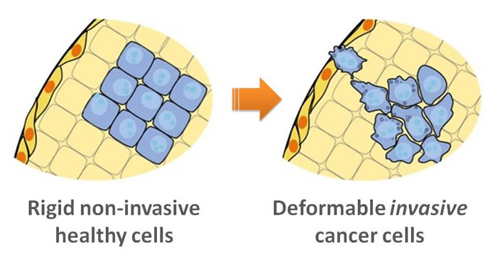
癌变细胞的变形能力要比正常细胞大得多。研究人员利用癌变细胞的这一特征开发出一种有多个小孔的微芯片,从... |

癌变细胞的变形能力要比正常细胞大得多。研究人员利用癌变细胞的这一特征开发出一种有多个小孔的微芯片,从胸水提取的细胞进入这些小孔后会撞上芯片的“墙壁”弹回而发生变形,变形程度会被高速成像设备记录下来,以每秒 100 个细胞的速度分析,从而判断是否存在癌细胞。

饶建宇指出,目前的癌症检查往往是间接地判断癌变细胞的一些行为特征,如浸润性和转移能力、对药物的敏感性等,一般要先对细胞进行固定处理再染色,或提取DNA及蛋白成分等进行分析,程序多而复杂,但所得结果往往是片面和间接的。
而微芯片技术则是直接判断癌变细胞的物理及行为特征,无需对细胞处理或染色,因此简单而快速,也更加精确。饶建宇表示:“这就好像判断一个人的角斗能力,光看高矮胖瘦或家庭背景等也许有一些帮助但不够,而直接的比赛是最管用的。人们谈癌色变往往是由于癌细胞具有浸润和转移的共性,同时又有千变万化的个性,因此以直接的方法来判断癌细胞的物理及行为特征尤为重要,这使得我们对癌细胞的认识更直接、全面和准确,对癌症的诊断由此上了一个新平台。” (转化医学网360zhyx.com)
原文检索:
Quantitative Diagnosis of Malignant Pleural Effusions by Single-Cell Mechanophenotyping
Biophysical characteristics of cells are attractive as potential diagnostic markers for cancer. Transformation of cell stateor phenotype and the accompanying epigenetic, nuclear, and cytoplasmic modifications lead to measureable changes in cellular architecture. We recently introduced a technique called deformability cytometry (DC) that enables rapid mechanophenotyping of single cells in suspension at rates of 1000 cells/s—a throughput that is comparable to traditional flow cytometry. We applied this technique to diagnose malignant pleural effusions, in which disseminated tumor cells can be difficult to accurately identify by traditional cytology. An algorithmic diagnostic scoring system was developed on the basis of quantitative features of two-dimensional distributions of single-cell mechanophenotypes from 119 samples. The DC scoring system classified 63% of the samples into two high-confidence regimes with 100% positive predictive value or 100% negative predictive value, and achieved an area under the curve of 0.86. This performance is suitable for a prescreening role to focus cytopathologist analysis time on a smaller fraction of difficult samples. Diagnosis of samples that present a challenge to cytology was also improved. Samples labeled as “atypical cells,” which require additional time and follow-up, were classified in high-confidence regimes in 8 of 15 cases. Further, 10 of 17 cytology-negative samples corresponding to patients with concurrent cancer were correctly classified as malignant or negative, in agreement with 6-month outcomes. This study lays the groundwork for broader validation of label-free quantitative biophysical markers for clinical diagnoses of cancer and inflammation, which could help to reduce laboratory workload and improve clinical decision-making.

来源:bio360
 腾讯登录
腾讯登录
还没有人评论,赶快抢个沙发