 腾讯登录
腾讯登录Nature:探秘骨骼的生长
| 导读 | 哈佛医学院的Kimberly Cooper和Seungeun Oh等人使用一种特殊的显微镜,观察了小鼠生长板处软骨细胞经历的改变,这种显微镜使他们可以定量活细胞的大小和密度。这项研究于今天发表在Nature杂志的网站上。
跳鼠jerboa有一双引人注目的双腿,这双腿比它的胳膊要长得多,能够帮助这种两条腿的沙漠啮齿类动物快速跳跃,以躲避它的天... |
哈佛医学院的Kimberly Cooper和Seungeun Oh等人使用一种特殊的显微镜,观察了小鼠生长板处软骨细胞经历的改变,这种显微镜使他们可以定量活细胞的大小和密度。这项研究于今天发表在Nature杂志的网站上。
跳鼠jerboa有一双引人注目的双腿,这双腿比它的胳膊要长得多,能够帮助这种两条腿的沙漠啮齿类动物快速跳跃,以躲避它的天敌。现在,这双腿也帮助科学家们进一步了解了骨骼的生长机制,揭示了一些影响骨骼生长的因素。这项研究最终将帮助科学家们治疗骨骼生长的缺陷。
生长板是位于骨骼生长末端的区域,当这一区域中的细胞增大增多时,骨骼就会伸长。这些细胞被称为软骨细胞,它们会形成软骨,为之后生长的成熟钙化骨提供支架。科学家们知道,骨骼生长的长度和速度取决于软骨细胞的数量和大小。但人们并不了解促使这些细胞生长的机制。
哈佛医学院的Kimberly Cooper和Seungeun Oh等人使用一种特殊的显微镜,观察了小鼠生长板处软骨细胞经历的改变,这种显微镜使他们可以定量活细胞的大小和密度。这项研究于今天发表在Nature杂志的网站上。
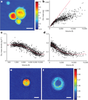
研究人员指出,小鼠的后腿生长时,软骨细胞经历了三个不同的阶段。首先,软骨细胞体积增大为三倍,同时细胞密度保持相对恒定。第二阶段,细胞显著膨胀,体积增至四倍,但细胞密度变小。第三阶段,细胞大小再次翻倍,但它们的密度保持不变。
研究人员在同一只小鼠中,对生长较快和生长较慢的骨骼进行了比较。他们发现,在生长较慢的前腿中,软骨细胞经过第一阶段的生长后,在第二阶段的中途停止生长。科学家们希望了解,与小鼠骨骼不同的近亲是否也采用类似的骨骼生长模式,于是又对埃及小跳鼠(Jaculus jaculus)的生长板进行了研究,他们发现,跳鼠胫骨的软骨细胞与小鼠生长模式类似,但跳鼠足部跖骨的生长模式却大不相同。研究显示,小鼠跖骨的软骨细胞在第三阶段只少量生长,但跳鼠的软骨细胞在这一阶段生长显著,体积达到原先的四十倍。
进一步研究显示,在小鼠后腿特异性地使胰岛素样生长因子1的编码基因失活,会导致软骨细胞在第二阶段后停止生长,胰岛素样生长因子1是一种影响生长和代谢的激素。研究表明,该基因可能在决定骨骼生长中具有关键作用,这一发现将有助于人们开发治疗骨骼生长缺陷的药物。
骨骼如何生长,这是一个老问题了。明确软骨细胞生长的三个阶段,将帮助科学家们进一步解析,引起各阶段变化的确切机制,康乃尔大学的Cornelia Farnum教授说。而这将帮助人们了解,不同形状骨骼的演化过程。
原文链接:
Multiple phases of chondrocyte enlargement underlie differences in skeletal proportions
The wide diversity of skeletal proportions in mammals is evident upon a survey of any natural history museum's collections and allows us to distinguish between species even when reduced to their calcified components. Similarly, each individual is comprised of a variety of bones of differing lengths. The largest contribution to the lengthening of a skeletal element, and to the differential elongation of elements, comes from a dramatic increase in the volume of hypertrophic chondrocytes in the growth plate as they undergo terminal differentiation1, 2, 3, 4, 5, 6, 7. However, the mechanisms of chondrocyte volume enlargement have remained a mystery8, 9, 10, 11. Here we use quantitative phase microscopy12 to show that mammalian chondrocytes undergo three distinct phases of volume increase, including a phase of massive cell swelling in which the cellular dry mass is significantly diluted. In light of the tight fluid regulatory mechanisms known to control volume in many cell types13, this is a remarkable mechanism for increasing cell size and regulating growth rate. It is, however, the duration of the final phase of volume enlargement by proportional dry mass increase at low density that varies most between rapidly and slowly elongating growth plates. Moreover, we find that this third phase is locally regulated through a mechanism dependent on insulin-like growth factor. This study provides a framework for understanding how skeletal size is regulated and for exploring how cells sense, modify and establish a volume set point.
来源:生物通
跳鼠jerboa有一双引人注目的双腿,这双腿比它的胳膊要长得多,能够帮助这种两条腿的沙漠啮齿类动物快速跳跃,以躲避它的天敌。现在,这双腿也帮助科学家们进一步了解了骨骼的生长机制,揭示了一些影响骨骼生长的因素。这项研究最终将帮助科学家们治疗骨骼生长的缺陷。
生长板是位于骨骼生长末端的区域,当这一区域中的细胞增大增多时,骨骼就会伸长。这些细胞被称为软骨细胞,它们会形成软骨,为之后生长的成熟钙化骨提供支架。科学家们知道,骨骼生长的长度和速度取决于软骨细胞的数量和大小。但人们并不了解促使这些细胞生长的机制。
哈佛医学院的Kimberly Cooper和Seungeun Oh等人使用一种特殊的显微镜,观察了小鼠生长板处软骨细胞经历的改变,这种显微镜使他们可以定量活细胞的大小和密度。这项研究于今天发表在Nature杂志的网站上。
研究人员指出,小鼠的后腿生长时,软骨细胞经历了三个不同的阶段。首先,软骨细胞体积增大为三倍,同时细胞密度保持相对恒定。第二阶段,细胞显著膨胀,体积增至四倍,但细胞密度变小。第三阶段,细胞大小再次翻倍,但它们的密度保持不变。
研究人员在同一只小鼠中,对生长较快和生长较慢的骨骼进行了比较。他们发现,在生长较慢的前腿中,软骨细胞经过第一阶段的生长后,在第二阶段的中途停止生长。科学家们希望了解,与小鼠骨骼不同的近亲是否也采用类似的骨骼生长模式,于是又对埃及小跳鼠(Jaculus jaculus)的生长板进行了研究,他们发现,跳鼠胫骨的软骨细胞与小鼠生长模式类似,但跳鼠足部跖骨的生长模式却大不相同。研究显示,小鼠跖骨的软骨细胞在第三阶段只少量生长,但跳鼠的软骨细胞在这一阶段生长显著,体积达到原先的四十倍。
进一步研究显示,在小鼠后腿特异性地使胰岛素样生长因子1的编码基因失活,会导致软骨细胞在第二阶段后停止生长,胰岛素样生长因子1是一种影响生长和代谢的激素。研究表明,该基因可能在决定骨骼生长中具有关键作用,这一发现将有助于人们开发治疗骨骼生长缺陷的药物。
骨骼如何生长,这是一个老问题了。明确软骨细胞生长的三个阶段,将帮助科学家们进一步解析,引起各阶段变化的确切机制,康乃尔大学的Cornelia Farnum教授说。而这将帮助人们了解,不同形状骨骼的演化过程。
原文链接:
Multiple phases of chondrocyte enlargement underlie differences in skeletal proportions
The wide diversity of skeletal proportions in mammals is evident upon a survey of any natural history museum's collections and allows us to distinguish between species even when reduced to their calcified components. Similarly, each individual is comprised of a variety of bones of differing lengths. The largest contribution to the lengthening of a skeletal element, and to the differential elongation of elements, comes from a dramatic increase in the volume of hypertrophic chondrocytes in the growth plate as they undergo terminal differentiation1, 2, 3, 4, 5, 6, 7. However, the mechanisms of chondrocyte volume enlargement have remained a mystery8, 9, 10, 11. Here we use quantitative phase microscopy12 to show that mammalian chondrocytes undergo three distinct phases of volume increase, including a phase of massive cell swelling in which the cellular dry mass is significantly diluted. In light of the tight fluid regulatory mechanisms known to control volume in many cell types13, this is a remarkable mechanism for increasing cell size and regulating growth rate. It is, however, the duration of the final phase of volume enlargement by proportional dry mass increase at low density that varies most between rapidly and slowly elongating growth plates. Moreover, we find that this third phase is locally regulated through a mechanism dependent on insulin-like growth factor. This study provides a framework for understanding how skeletal size is regulated and for exploring how cells sense, modify and establish a volume set point.
来源:生物通
还没有人评论,赶快抢个沙发